
Scientists Identify Unusual Brain Waves Linked to Memory Loss
Guest Contributor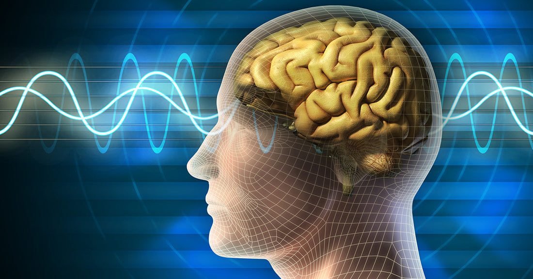
Picture walking out of a grocery store at dusk, bags in hand, only to stop short in the parking lot and wonder: where exactly did you leave your car? That brief, disorienting lapse is a tiny example of episodic memory at work, or in this case, not quite working. A new study from the University of California, Berkeley, digs deeply into why these kinds of memories fade with age and points to an unexpected culprit hiding in our sleeping brains: disrupted deep sleep brain waves linked to Alzheimer’s related tau protein.
Episodic memory allows people to recall specific personal experiences, from the first day of school to a conversation that took place an hour ago. Researchers have long known that this form of memory tends to weaken as people get older. At the same time, they know that memory decline can become far more severe in Alzheimer’s disease. What has remained elusive is exactly what begins to go wrong inside the brain long before full-blown symptoms appear. The new work, published in Nature Neuroscience by neuroscientist Omer Sharon and colleagues under the direction of sleep researcher Matthew Walker, suggests that early changes in deep sleep may be a critical part of the story.

The team focused on non-REM deep sleep, a stage in which dreaming is limited but the brain performs crucial housekeeping and memory related tasks. During this phase, large populations of neurons repeatedly switch off and on together in a highly synchronized way. These coordinated shifts generate slow electrical waves that ripple across the brain, typically beginning in the frontal cortex. Sharon describes each slow wave as reflecting an enormous population of neurons shutting down and restarting in unison, with the sequence cascading across wide brain regions.
This elegant choreography appears vital for memory consolidation overnight, the process through which fragile new experiences are stabilized and stored. The study suggests that the more smoothly these slow waves travel across the brain, the better the brain preserves newly learned information. I found this detail striking because it ties an abstract idea like “sleep quality” to something concrete and measurable: how far and how coherently these slow waves move.
The researchers discovered that in older adults, this coordinated movement often breaks down. Instead of broad, traveling waves spanning distances equivalent to the width of a hand on the scalp, the slow waves in older brains tend to travel shorter distances and appear more independently. In other words, instead of a unified wave sweeping across regions, the patterns become fragmented and scattered. The team linked this disruption to both memory performance and the buildup of tau, a protein strongly associated with Alzheimer’s pathology.
To observe these sleep-related changes, the scientists recorded brain activity during sleep using electroencephalograms, or EEGs. They compared a group of cognitively healthy young adults in their early twenties with older adults in their mid-sixties to mid-seventies. The contrast was clear. Younger participants showed smooth, large-scale traveling slow waves. Older participants showed shorter range waves that often popped up in isolation rather than as part of a flowing sequence. These altered patterns were not just curiosities in the data. They lined up with measurable differences in how well people retained information overnight.
To connect these sleep signatures to Alzheimer’s related changes, the team worked with imaging specialist William Jagust to perform PET scans on the older participants. PET imaging uses radioactive tracers in the bloodstream to visualize biological processes, including the distribution of tau protein in the brain. The scans revealed that greater tau accumulation in the frontal cortex correlated with a breakdown in those long-distance traveling slow waves that are typical of deep, restorative sleep.
Importantly, these older adults did not have diagnosed Alzheimer’s disease. Their memory scores fell within the normal range for their age, yet they already showed tau buildup and disrupted sleep waves. This highlights a key point. The study suggests that Alzheimer’s related changes, such as tau in frontal brain regions, can be present well before clinical symptoms of dementia appear. Even within the “normal” range of aging, the amount and location of tau tracked with more subtle declines in cognitive performance and with changes in sleep architecture.
The team then took a closer look at how these sleep patterns related to actual memory outcomes. Participants learned pairs of associated words in the evening, then returned the next day to see how many pairs they could recall. People whose slow waves traveled shorter distances and appeared more isolated remembered fewer word pairs. Sharon and colleagues labeled these disconnected patterns “lonely waves,” a phrase that vividly captures the idea of brain activity that fails to join a broader, cooperative network.
The researchers also followed some participants over several years. Those who showed rising levels of tau in the frontal cortex over time also showed a progressive breakdown in coordinated slow waves. At the same time, their ability to retain newly learned information across a night of sleep declined. The findings do not prove that tau directly causes disrupted waves or that the waves directly cause memory loss, but they do show that tau accumulation, lonely waves, and worsening episodic memory tend to climb together.

To broaden the evidence, the group collaborated with neurologist Yo-El Ju at Washington University in St. Louis and analyzed spinal fluid from another group of older adults. This second method did not pinpoint tau in specific brain regions the way PET scans do. Instead, it provided overall levels of tau relative to amyloid, another protein closely tied to Alzheimer’s disease. Even so, the pattern looked familiar. People whose EEG recordings showed more isolated slow waves during sleep had higher ratios of tau to amyloid in their spinal fluid. This consistency across different participants and techniques added weight to the notion that certain deep sleep disruptions are tightly linked to Alzheimer’s related protein changes.
The emerging picture is that deep sleep, and particularly the integrity of traveling slow waves, may offer an early window into brain health. Memory decline in aging is often described as inevitable, yet this study suggests that it may be influenced by how much underlying pathology accumulates in frontal regions that generate global slow waves. As Sharon notes, it may not simply be about chronological age, but about how much tau is present in the areas where those widespread waves originate.
These insights raise intriguing possibilities. In the future, researchers might be able to use specific sleep patterns as early indicators of risk for memory problems or to evaluate whether interventions are helping preserve brain function. They also point toward sleep itself as a possible target for strategies that aim to support healthy aging. At the same time, a central question remains unresolved. It is not yet clear whether tau buildup drives the disruption of sleep waves, whether impaired sleep promotes tau accumulation, or whether a third set of processes influences both.
For now, the concept of “lonely waves” offers a compelling metaphor for what may be happening inside the aging brain. Instead of vast networks of neurons working together during deep sleep to preserve the day’s experiences, some brains appear to slip into more fragmented, isolated patterns. As researchers continue to track how tau, sleep, and memory change over time, their findings may help inform future approaches to diagnosing and treating age-related cognitive decline. Read more at https://www.sciencedaily.com/releases/2026/11/261101010556.htm